The 7 Types of Eczema: A Complete Guide to Identifying and Treating Each
“Eczema” is not a single diagnosis. It is an umbrella term for a group of conditions that leave skin inflamed, itchy, and dry. Dermatologists recognize seven main types, and while they all share that core picture, they differ in where they appear, what sets them off, what they look like up close, and how they are treated.
Putting the right name to a rash matters, because management varies a great deal from one type to the next. The barrier creams and bleach baths that calm atopic dermatitis do little for the circulation problem driving stasis dermatitis. The antifungal shampoo that clears seborrheic dermatitis is the wrong tool for a coin of nummular eczema. Identifying the type is the first real step toward relief.
This guide walks through all seven side by side: what each one looks like, where it tends to show up, what triggers it, and how treatment differs. One caution before we start. These types overlap, one person can have more than one at the same time, and several of them mimic other skin diseases such as ringworm, psoriasis, and infection. Only a board-certified dermatologist can give you a firm diagnosis. Use this as a map, not as a substitute for that visit.
1. Atopic dermatitis
Atopic dermatitis is the most common type, and it is the one most people mean when they simply say “eczema.” It usually begins early in life, often between 2 months and 5 years of age, though it can start during puberty or adulthood. It is a chronic, relapsing condition rooted in a weakened skin barrier (often tied to filaggrin gene mutations) and an overactive immune response. It frequently travels with asthma, hay fever, and food allergy as part of the atopic march.
What it looks like: itchy, dry, inflamed skin. In babies it favors the cheeks and scalp. On lighter skin it tends to look pink or red, while on darker skin it more often appears as brown, gray, or purple patches with small bumps and intense dryness.
Where it shows up: in infants, the face and the outsides of the limbs. In children and adults, it moves to the flexural areas, meaning the creases inside the elbows and behind the knees, plus the neck, wrists, ankles, and hands. Severe, raw patches can ooze, a problem known as weeping eczema.
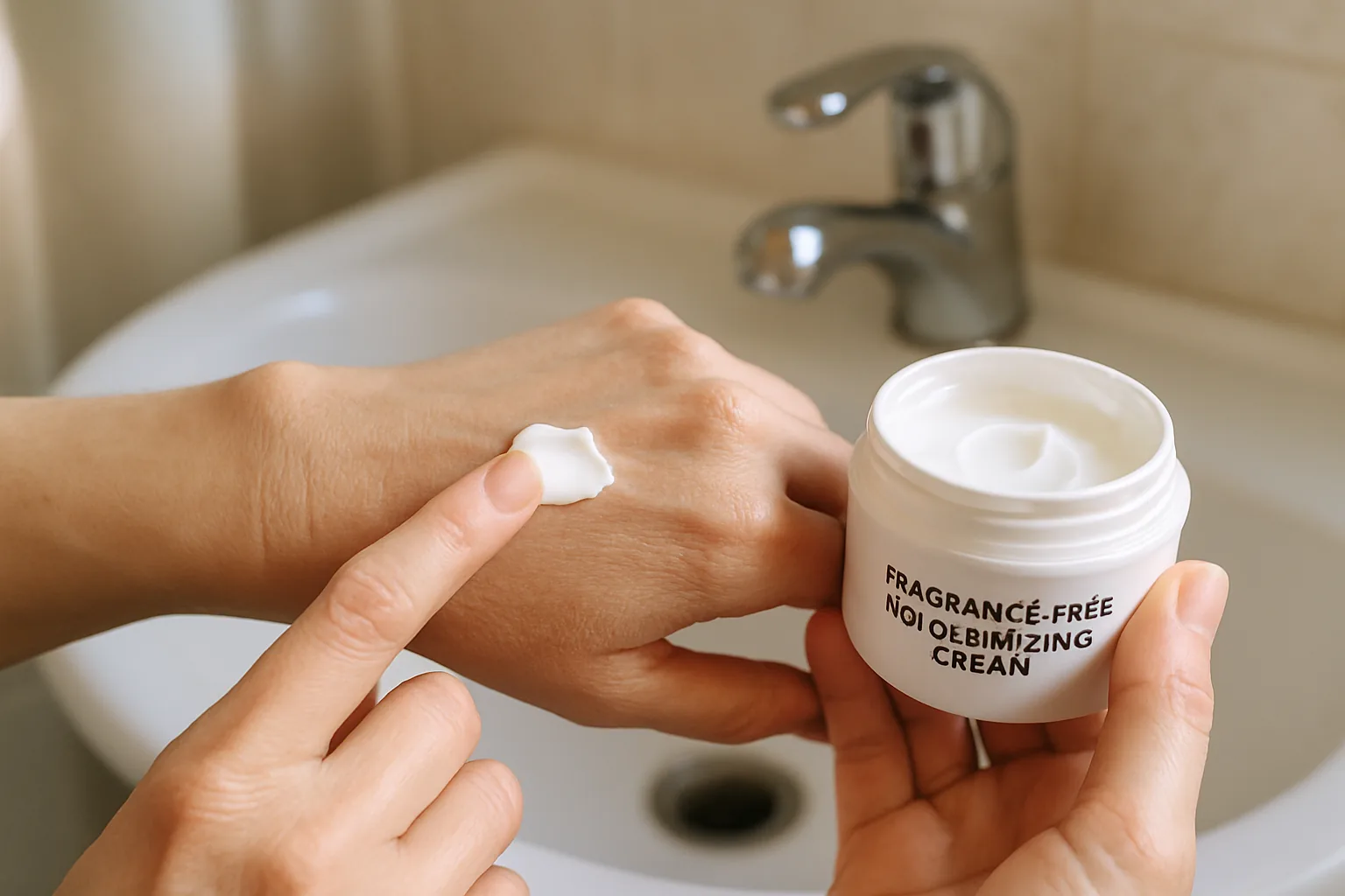
How it is managed: the foundation is daily barrier care, a fragrance-free emollient (ceramide-based creams work well) applied with the soak and seal method, plus gentle cleansers. Topical corticosteroids calm flares, while calcineurin inhibitors (tacrolimus, pimecrolimus) and the PDE4 inhibitor crisaborole offer steroid-sparing options. Moderate to severe disease may call for biologics such as dupilumab or newer JAK inhibitor therapies.
2. Contact dermatitis
Contact dermatitis is a reaction to something that touches the skin. It comes in two forms. Irritant contact dermatitis, the more common one, is direct damage from substances like soaps, solvents, and frequent water exposure. Allergic contact dermatitis is an immune reaction to a specific allergen. According to the American Academy of Dermatology, more than 15,000 substances can trigger it, with nickel and fragrance among the most common offenders.
What it looks like: an itchy red or discolored rash, sometimes with blisters, usually confined to the exact area that touched the trigger. A telltale clue is a rash in the shape of a watchband, a necklace, or a waistband. With the allergic form, the rash can appear hours or even days after contact.
Where it shows up: the hands are a classic site, especially in healthcare workers, hairdressers, and anyone who does frequent wet work. The eyelids and face are common too, often from cosmetics or nail products carried up by the fingers.
How it is managed: the only true cure is finding and avoiding the culprit, which is why patch testing is so valuable for the allergic form. Barrier protection (gloves, fragrance-free cream after every wash) helps, and topical steroids settle active flares. Stubborn hand involvement can be one of the hardest forms to control, and the options are expanding. See our explainer on a new topical for chronic hand eczema for where the research is heading.
3. Dyshidrotic eczema
Dyshidrotic eczema, also called pompholyx, announces itself with sudden crops of tiny, deep-seated, intensely itchy blisters. They are often compared to tapioca pearls sitting just under the skin.
What it looks like: clusters of small fluid-filled blisters on the palms, the soles, and the sides of the fingers and toes. As they resolve, the skin tends to peel, crack, and feel painfully dry.
Where it shows up: the hands and feet, almost exclusively. It is most common in adults between 20 and 40, and somewhat more frequent in women.
How it is managed: high-potency topical steroids during a flare, cool compresses, and careful soaking and drying. Because sweat, stress, hot weather, and nickel or cobalt sensitivity are frequent triggers, sweat control and allergy evaluation help. Severe, recurrent cases may need phototherapy or systemic medication. Since the hands are so often involved, much of the hand eczema treatment landscape applies here too.
4. Nummular eczema
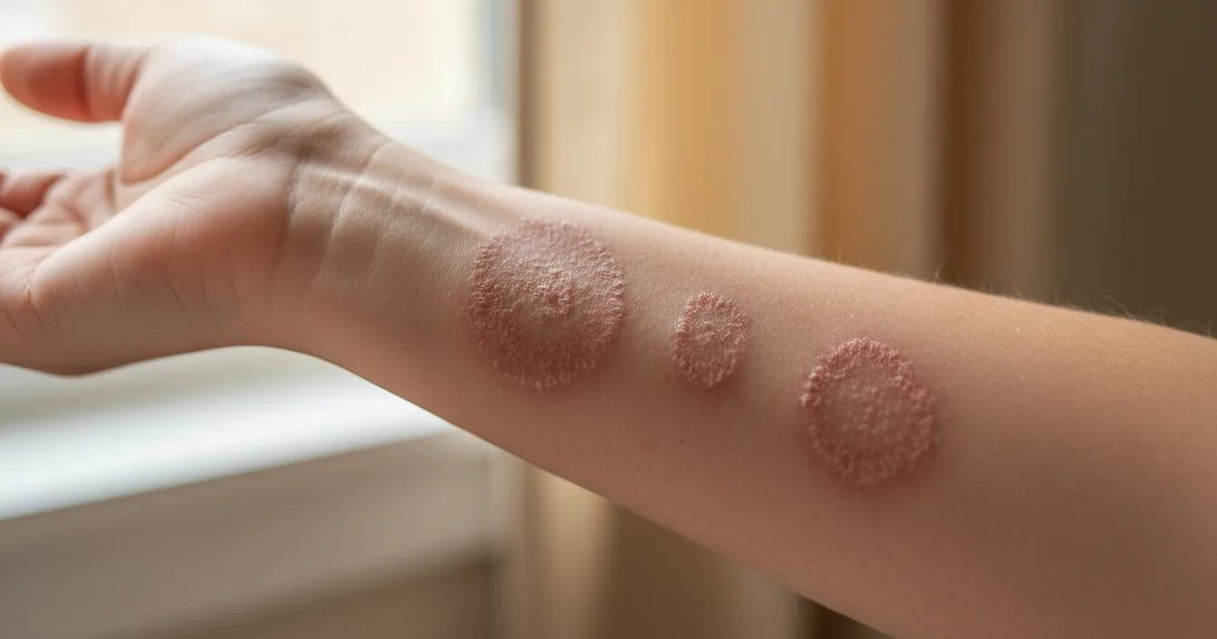
Nummular eczema takes its name from the Latin word for coin, and the shape is its signature. It is also called discoid eczema.
What it looks like: distinct round or oval patches, anywhere from a coin to a few inches across. They can be dry and scaly or wet, oozing, and crusted, and they are often intensely itchy or burning. Because the patches are so well defined, nummular eczema is frequently mistaken for ringworm or psoriasis.
Where it shows up: most often the lower legs, arms, hands, and torso. It commonly follows a skin injury such as an insect bite, a scrape, or a burn, and it is strongly linked to very dry skin.
How it is managed: generous moisturizing is essential, paired with mid to high-potency topical steroids. These patches readily become colonized with staph bacteria, so dermatologists watch closely for infection and treat it when present. The condition can be stubborn and prone to returning, so consistent barrier care between flares pays off.
5. Seborrheic dermatitis
Seborrheic dermatitis settles in the oil-rich zones of the skin. In infants it is the familiar cradle cap, and in adults its mildest form is everyday dandruff.
What it looks like: greasy yellow or white flaky scale sitting on red or discolored skin. It can itch, though often less fiercely than other types.
Where it shows up: the scalp, eyebrows, the sides of the nose, behind and inside the ears, the central chest, and skin folds. It tends to favor exactly the places where oil glands are most active.
How it is managed: the condition is linked to an overgrowth of Malassezia yeast on the skin, not to poor hygiene, so antifungal treatment is the backbone. Medicated shampoos with ketoconazole, selenium sulfide, or zinc pyrithione help on the scalp, and topical antifungals or short courses of mild steroids or calcineurin inhibitors calm the face. It is chronic and tends to flare with cold weather, stress, and illness, so it is managed over time rather than cured outright.
6. Stasis dermatitis
Stasis dermatitis, also known as gravitational or venous eczema, is the one type driven mainly by a circulation problem rather than by an allergy or barrier defect.
What it looks like: swelling, redness or a brownish discoloration, scaling, and itch on the lower legs and ankles. Over time the skin can thicken and harden, and advanced cases may develop open sores called venous ulcers.
Where it shows up: the lower legs, almost always. It is most common in older adults and in people with varicose veins, high blood pressure, heart or kidney conditions, or a history of blood clots and leg swelling. When the valves in the leg veins weaken, blood and fluid pool and leak into the surrounding skin, setting off inflammation.
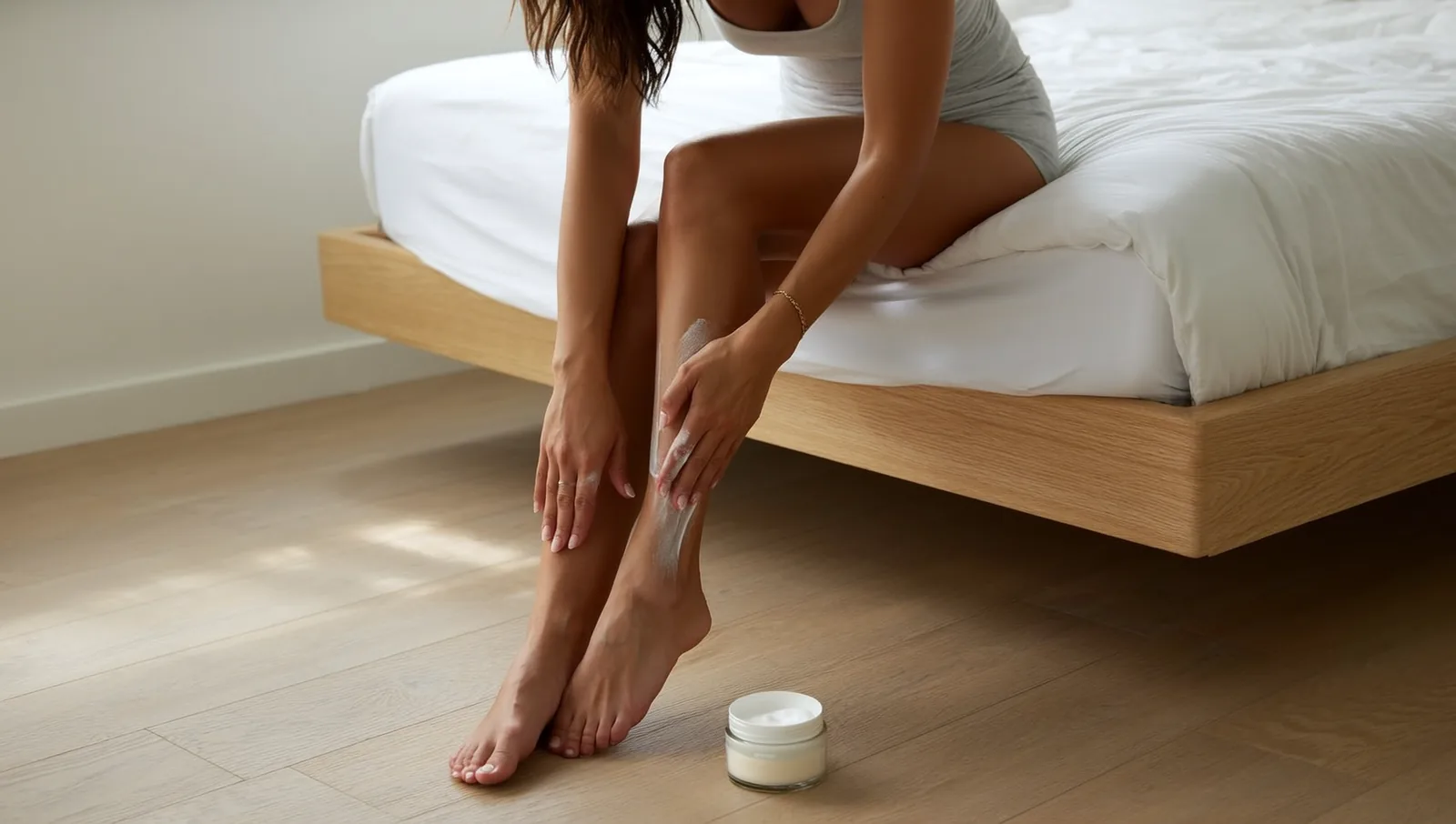
How it is managed: treating the underlying circulation is the priority. Compression stockings, regular leg elevation, and walking to work the calf muscles all reduce the pooling. Moisturizers protect the fragile skin, topical steroids quiet active flares, and any sign of infection or ulceration needs prompt medical care. Self-care is a genuine cornerstone of getting this type under control.
7. Neurodermatitis
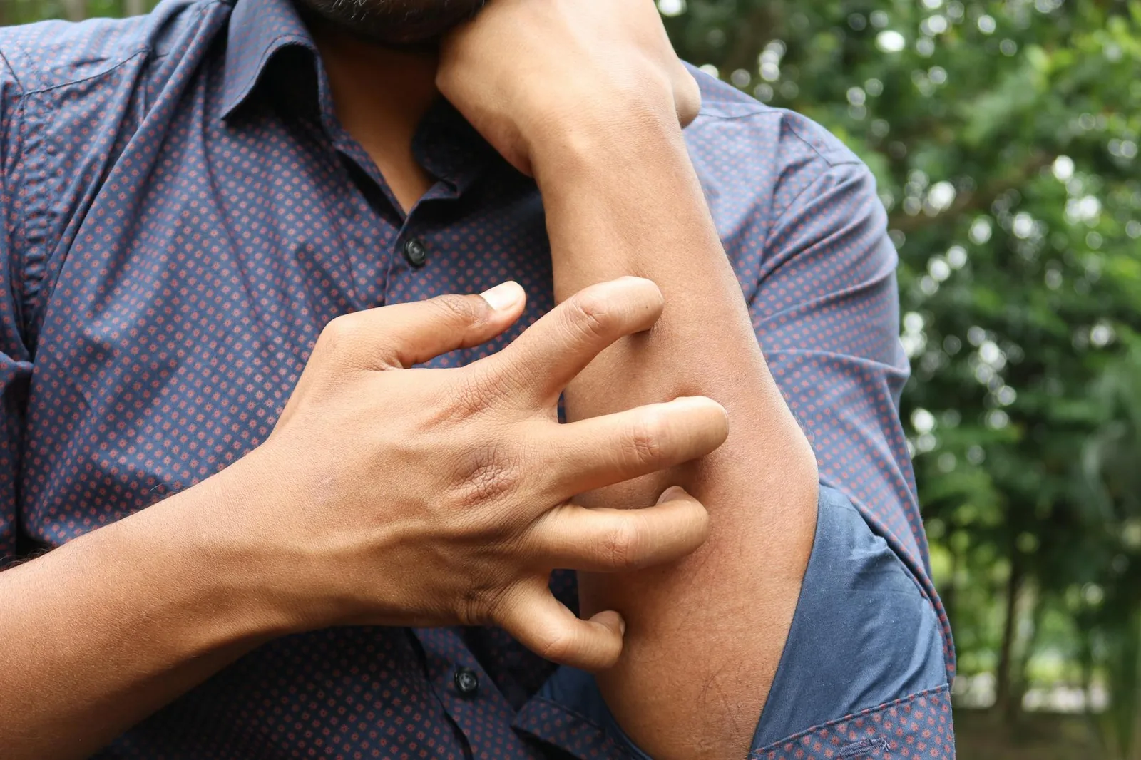
Neurodermatitis, also called lichen simplex chronicus, begins with an itch and is sustained by the response to it.
What it looks like: one or a few thickened, leathery patches where the normal skin lines look exaggerated, a change dermatologists call lichenification. The patches can crack, bleed, or become infected from repeated scratching.
Where it shows up: the ankles, the nape of the neck, the scalp, the wrists and forearms, and the genital or anal area. People often report that the itch is worst when they are relaxing or just before sleep.
How it is managed: the goal is to break the itch-scratch-itch cycle. That means physically interrupting the scratching (covering or occluding the patch), high-potency topical steroids that are sometimes applied under occlusion, and antihistamines to ease nighttime itch. Because the condition frequently begins during a stressful period, addressing stress and anxiety is part of real treatment. It rarely clears on its own.
The seven at a glance
When you are trying to narrow things down, the standout clue usually points the way:
- Itchy rash in the elbow and knee creases since childhood, with a personal or family history of allergies: atopic dermatitis
- A rash exactly where something touched the skin: contact dermatitis
- Crops of tiny, deep itchy blisters on the palms or soles: dyshidrotic eczema
- Round, coin-shaped patches: nummular eczema
- Greasy yellow scale on the scalp, eyebrows, or sides of the nose: seborrheic dermatitis
- Swollen, discolored, itchy lower legs: stasis dermatitis
- One thick, leathery patch you cannot stop scratching: neurodermatitis
Getting the right diagnosis
Because these types overlap and several of them imitate other conditions, self-diagnosis only goes so far. Eczema and psoriasis in particular can look alike to an untrained eye, yet they are treated quite differently. A dermatologist has tools to sort this out, including patch testing for suspected contact allergy and a simple skin scraping to rule out a fungal infection that might be masquerading as nummular eczema.
A few principles apply across every type. Never stop a prescribed medication without talking to your doctor first. Watch for signs of secondary infection, since broken, scratched, or weeping skin is vulnerable to bacteria: increasing redness, warmth, swelling, yellow crusting or pus, and fever all warrant prompt medical attention. And remember that eczema is highly individual. A routine that transforms one person’s skin may do little for another, so expect some trial and error as you and your dermatologist find your combination.
The bottom line
Seven types, one shared thread of inflamed and itchy skin, but seven different maps and seven different toolkits. Knowing whether you are dealing with the childhood-onset barrier problem of atopic dermatitis, the trigger-driven rash of contact dermatitis, the blistering of dyshidrotic eczema, the coins of nummular eczema, the greasy scale of seborrheic dermatitis, the circulation-linked swelling of stasis dermatitis, or the scratch-driven patches of neurodermatitis changes what you do next. If your skin has been flaring without a clear answer, that clarity is worth a visit to a board-certified dermatologist.
Further reading (sources)
- American Academy of Dermatology on the major types of eczema and how they differ
- American Academy of Dermatology for an overview of atopic dermatitis
- National Eczema Association for a patient-friendly tour of the eczema types
- Medscape on the pathophysiology behind atopic dermatitis
- National Eczema Association on the types of eczema and how they differ